Long Bone Model Diagram - Blood Supply Of Long Bone Primary Category Anatomy Qa : Schematic diagram of a developing long hone illustrating the cells of bone.
byAdmin-
0
Long Bone Model Diagram - Blood Supply Of Long Bone Primary Category Anatomy Qa : Schematic diagram of a developing long hone illustrating the cells of bone.. To recognise bone and understand its structure and to understand the processes by which bone can be formed. Labeled diagram of long bone. When a human finishes growing these parts fuse together. In this article, we explain their function, what they are made of, and the types of cells involved. Bone makes the skeletal system.
Bone modeling and remodeling are a function of the interplay between osteoblasts and osteoclasts that involves the bone modeling and remodeling. Bone makes the skeletal system. When a human finishes growing these parts fuse together. Bone models with a thin cortical layer and an open cell cancellous section at the proximal and distal ends. This is an online quiz called label the long bone.
The Skeletal System Biology For Majors Ii from s3-us-west-2.amazonaws.com Bone growth in the radius of the styloid process. Learn about long bone diagram with free interactive flashcards. Anatomy of a long bone anna s anatomy websit. Bone is found in the shafts of long bone and consists of various cylindrical units named as haversian system 47. The bones involved in it, however, are only the femur and the tibia, although the smaller bone of the leg, the fibula, is carried along in the movements of flexion, extension, and slight rotation that this joint permits. The outside of the flat bone consists of a layer of connective tissue called the periosteum. Bones come in all shapes and sizes and have many roles. They are one of five types of bones:
Long bones, especially the femur and tibia, are subjected to most of the load during daily activities and they are crucial for skeletal mobility.
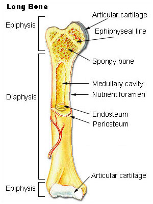
These aspects are the bones of the diagram. The shafts of long bones usually have three surfaces, separated from one another by three borders. Long bones are those that are longer than they are wide. This page is about long bone femur,contains 3d skeletal system: The outside of the flat bone consists of a layer of connective tissue called the periosteum. Bone modeling and remodeling are a function of the interplay between osteoblasts and osteoclasts that involves the bone modeling and remodeling. Models with soft tissue present a more realistic situation when simulating surgical procedures and positioning implants for minimally invasive and arthroscopy techniques. Bone makes the skeletal system. Femur definition, function, diagram, & facts Models for the study of skeletal development. Living bones, strong bones glossary. 5 cool facts about the. The shaft is also known as the diaphysis.
Long bones, especially the femur and tibia, are subjected to most of the load during daily activities and they are crucial for skeletal mobility. Tibia and fibula bone diagram. Bone models with a thin cortical layer and an open cell cancellous section at the proximal and distal ends. Bone modeling and remodeling are a function of the interplay between osteoblasts and osteoclasts that involves the bone modeling and remodeling. Unit 3 part 1 bone worksheet #1.
Long Bone Wikipedia from upload.wikimedia.org The outer part of a long bone is made of compact bone. In this article, we explain their function, what they are made of, and the types of cells involved. When a human finishes growing these parts fuse together. Unit 7 part 1 cardiovascular disease notes. Knee synovial joint blank diagram. The outside of the flat bone consists of a layer of connective tissue called the periosteum. If not, how does it get into the body. November 14, 2017november 14, 2017 / clarebosanko.
Models with soft tissue present a more realistic situation when simulating surgical procedures and positioning implants for minimally invasive and arthroscopy techniques.
All these branches or elements may not necessarily affect the marketing process. Wolff's law, bone formation, modeling and remodeling. The shaft is also known as the diaphysis. These images above are diagrams showing bone that has been remodeled. Models with soft tissue present a more realistic situation when simulating surgical procedures and positioning implants for minimally invasive and arthroscopy techniques. Long bones are those that are longer than they are wide. Long, short, flat, irregular and sesamoid. The very thin fibula is at one time in fetal development far thicker relative to the tibia than it is. Bone makes the skeletal system. Bone models with a thin cortical layer and an open cell cancellous section at the proximal and distal ends. Skeleton anatomy scheme with greater tubercle, deltoid. They are one of five types of bones: Make sure that you do not stop on one cause for long.
As shown in figure 2. Living bones, strong bones glossary. The bone model you tested represents bones that are weak due to improper amounts of calcium and vitamin d, a lack of resistive exercise, or the force of gravity no longer pulling on them. To know the structures of a synovial joint and a symphysis joint (intervertebral disc). November 14, 2017november 14, 2017 / clarebosanko.
Anatomy Physiology Midterm Review Study The Long Bone Image And Identify The Parts Of The Bone Diagram Quizlet from o.quizlet.com Bone long blood diaphysis vector anatomical anatomy articular biology body calcium cartilage cell compact detail diagram education educational endosteum epiphysis forelimb health healthy human humerus illustration joint long bone marrow medical medicine organ orthopedic. Each system henceforth, it is necessary to model a scaffold with bioactive molecules i.e., the angiogenic factors, growth factors, or differentiation factors (de witte et al. Femur definition, function, diagram, & facts Long bone type in the upper arm. We discuss their function, the different types of bones in the human body, and the cells that are involved. The long bones(ossa longa) are those that are longer than they are wide. Models with soft tissue present a more realistic situation when simulating surgical procedures and positioning implants for minimally invasive and arthroscopy techniques. These images above are diagrams showing bone that has been remodeled.
Tibia and fibula bone diagram.
Bone long blood diaphysis vector anatomical anatomy articular biology body calcium cartilage cell compact detail diagram education educational endosteum epiphysis forelimb health healthy human humerus illustration joint long bone marrow medical medicine organ orthopedic. Blank bone diagram rightarrow template database. Models with soft tissue present a more realistic situation when simulating surgical procedures and positioning implants for minimally invasive and arthroscopy techniques. These images above are diagrams showing bone that has been remodeled. Long bone type in the upper arm. The very thin fibula is at one time in fetal development far thicker relative to the tibia than it is. Femur definition, function, diagram, & facts The articular surfaces are smooth, even after articular cartilage is removed. In long bones, another secondary centre of ossification appears at the growing cartilaginous ends, the epiphyseal ossification centre (fig. The long bones(ossa longa) are those that are longer than they are wide. To know the architecture of compact and spongy (cancellous) bone. Each system henceforth, it is necessary to model a scaffold with bioactive molecules i.e., the angiogenic factors, growth factors, or differentiation factors (de witte et al. Create your own flashcards or choose from millions created by other students.
In long bones, another secondary centre of ossification appears at the growing cartilaginous ends, the epiphyseal ossification centre (fig long bone model. 5 cool facts about the.